
Iron-Stained vs Iron-Included Quartz
A careful field guide to locating red-brown colour in quartz: exterior deposit, surface-connected fracture material or an apparently enclosed feature—without turning visual clues into laboratory identification.
In this field note
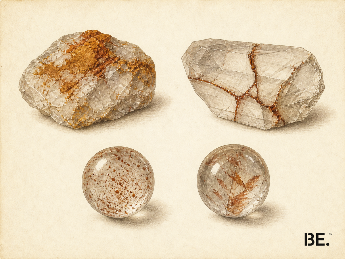
“Stained” and “included” sound like opposite labels. In practice, quartz presents a third case between them: material can enter or form along a fracture connected to the exterior and remain visible well inside a crystal or bead. Describing where the colour sits is therefore safer than naming what it is or when it formed.
This guide is an observation method, not a home identification test. It keeps surface evidence, fracture geometry and apparently enclosed features separate, then marks the point at which laboratory work becomes the honest next step.
Why stain and inclusion are not a simple binary
In gemmology, an internal feature may be older than its host, form with it, or arrive later. Those relationships are often described as protogenetic, syngenetic and epigenetic. The vocabulary is useful, but the timing is not always recoverable from a photograph. A feature that appears “inside” may follow a fracture with an opening outside the field of view.
GIA has documented iron-hydroxide debris along extensive surface-reaching fractures in rose quartz. It has also documented identifiable hematite platelets enclosed in quartz. Both can be visible beneath a polished surface; they are not the same evidence pattern. The defensible first question is where does the feature run?, not which mineral name does the colour suggest?
Three places colour can sit
| Observed position | Plausible explanations | What the observation does not prove |
|---|---|---|
| Exterior surface | A later mineral deposit, polishing or cleaning residue, environmental dirt, or a surface treatment. | Mineral species, natural versus applied origin, or the history of the whole stone. |
| Surface-connected fracture | A later fluid-borne deposit, natural fracture fill, treatment material, dye or residue entering an open path. | That the material grew with the quartz, or that it is natural or artificial. |
| Apparently enclosed feature | A solid or fluid feature trapped in the host, a feature of another growth stage, or a connection to the exterior that is not visible. | Exact species, age relationship, locality or treatment history. |
Depth cues matter. A truly surface-bound patch stays attached to the outer contour as the piece turns. A deeper feature may show parallax against the far surface. A fracture-bound deposit often follows a planar or branching line and may be traceable towards an edge, pit or drill hole. None of these clues is exclusive: polish, curvature and lighting can imitate depth.
How to observe without overclaiming
- Record before cleaning. Photograph the surface and any residue as received. If loose dust prevents viewing, use only a soft dry cloth after recording it. Do not begin with acid, rust remover, abrasion or an ultrasonic cleaner on a finished piece.
- Use diffuse light. Record the colour distribution without glare.
- Add transmitted light. If the piece allows light through, look for depth, fracture planes and continuity.
- Add oblique reflected light. Low-angle light can reveal whether material sits in a pit, on polish or along a surface break.
- Rotate, do not just zoom. Parallax is more informative than a single enlarged frame.
- Trace the feature. Check whether it reaches an edge, a drill hole, a pit or a visible fracture opening.
- Write the observation before the interpretation. “Red-brown material along a surface-reaching fracture” is stronger evidence language than “natural hematite inclusion”.
A 10× loupe can improve the view, but it does not change the evidence category. It may reveal geometry that supports one hypothesis over another; it does not turn colour into chemical analysis.
What photographs cannot prove
Mineral species
Red, orange and brown appearances are not exclusive to hematite. Other minerals, alteration products, residues, dyes and lighting conditions can overlap visually. Even a familiar platelet or needle shape is a candidate, not a species result.
Formation timing
Material along a sealed-looking line may have entered through a fracture before polishing. An apparently free-standing feature may belong to an earlier, simultaneous or later event. Without a complete three-dimensional path and suitable analysis, the timing remains unresolved.
Origin and treatment
Neither a red-brown colour nor an internal-looking feature establishes a mine, country, natural origin or untreated status. Disclosure and laboratory evidence answer different questions from a visual location check. The BE. treatment disclosure chart keeps identity and treatment on separate lines.
When laboratory testing matters
Escalate when the mineral name, treatment, geographic origin or value changes the buying, insurance or resale decision. Gemmological laboratories may combine microscopy with Raman spectroscopy, X-ray diffraction or other methods according to the question. No single method answers every question, and a result belongs to the tested object rather than every visually similar bead.
For ordinary purchasing, ask for a precise material description and treatment disclosure. If the seller's conclusion rests only on colour, request the evidence behind it. If uncertainty remains, retain the neutral observation instead of upgrading it into a claim.
Relationship to hematoid quartz
The Hematoid Quartz guide covers the named material, its iron-oxide context and its quality language. The iron and crystal colour guide addresses the wider question of why iron can produce different colours.
Frequently asked questions
Q1.Is iron-stained quartz the same as iron-included quartz?
Not necessarily. Colour may sit on the exterior, line a surface-connected fracture, or appear in an enclosed feature. A fracture deposit can look internal while still being epigenetic.
Q2.Can a photograph prove whether the colour is a stain or an inclusion?
No. Lighting, polish, fracture geometry and an unseen surface connection can change the appearance. A photograph can guide closer observation, not establish mineral identity or formation history.
Q3.Does red-brown colour prove hematite?
No. Hematite is one possibility, but colour alone does not distinguish it from other minerals, alteration products, treatments or residues. Identification requires suitable testing.
Q4.If colour follows a fracture, is it still ‘inside’ the quartz?
It may be visible inside the bead, but the fracture can connect to the surface and carry a later deposit. That is different from proving a growth inclusion by sight.
Q5.Should I use acid or stain remover on finished quartz jewellery?
Not as a first diagnostic step. Chemicals can affect metal, cord, coatings, fracture fillings or the evidence you are trying to examine. Ask a qualified professional before treating a finished piece.
Q6.Is surface staining automatically poor quality?
No. Quality depends on accurate disclosure, stability, finish and the intended appearance. A surface feature is evidence to describe, not an automatic grade.
References
- GIA: Colored Stones Unearthed
- GIA: A Shrinkage “Footprint” in Rose Quartz
- GIA: Hematite in Quartz
- GIA, Gems & Gemology Fall 2013: Epigenetic Malachite in Quartz
- Applied Geochemistry: Iron-oxide precipitates on quartz surfaces
- GIA: How GIA Analyses Coloured Stones
- GIA: Raman Spectroscopy and X-Ray Diffraction
Field Notes.
One stone at a time: its geology, and how to read quality. The Stone Buying Checklist comes with your first email.
By subscribing you agree to receive emails from BE. Unsubscribe anytime.
Sent. Check your inbox.