Why Iron Looks Red Sometimes — and Green, Blue or Yellow Other Times
Aquamarine is blue because trace iron behaves differently inside beryl. A material-first note on Fe2+, beryl, heat treatment and choosing blue crystal jewellery.
In this field note
The first time you notice this, it is genuinely strange. Garnet is iron-rich and reads deep red. Aquamarine is also iron-bearing and reads sky-blue. Both contain iron. Both contain a lot of it. They look nothing alike. If iron has a colour, why does it keep changing its mind?
The short answer is that iron has no single colour. What we call a stone’s colour is not the colour of an element sitting inside the lattice. It is the colour the host structure permits an electron transition to take, given the oxidation state of iron and the geometry of the site it occupies. Change any one of those variables and the visible result changes with it.
Two oxidation states, two electron worlds
Iron exists in nature as two stable cations: Fe²⁺ (ferrous, four 3d electrons after pairing) and Fe³⁺ (ferric, five). Their absorption behaviour is not similar. Fe²⁺ absorbs in a broad region around 1,000 nm and into the near-infrared, leaving relatively narrow paths through the visible band; Fe³⁺ has spin-forbidden bands across much of the visible. Each gives a fundamentally different palette.
The redox state is set during formation. Reducing conditions (low oxygen, deep crust, hydrothermal fluids near serpentinites) favour Fe²⁺. Oxidising conditions (surface weathering, late-stage hydrothermal alteration, magma exposed to atmospheric oxygen) push iron to Fe³⁺. The same iron atom can move between the two states across a stone’s history, but at the moment colour locks in, only one is dominant.
Coordination geometry: where iron sits matters
Iron substitutes into mineral structures by replacing other cations of similar size — usually Mg²⁺, Al³⁺, or Ca²⁺. The site iron occupies has a specific geometry: tetrahedral (4 surrounding oxygens), octahedral (6), or larger 8-fold coordination. The geometry controls the energy gap between d-orbitals, which controls which wavelengths are absorbed.
This is the part most explanations skip. Two minerals can hold the same iron in the same oxidation state and still look different because the bond distances and angles around that iron are different. Beryl’s six-fold beryllium ring puts Fe²⁺ in a channel with one set of distances; olivine’s octahedral M-sites give different distances; garnet’s eight-fold dodecahedral site is different again. Each site reshapes the d-orbital splitting and shifts the visible absorption.
Intervalence charge transfer: a third lever
When Fe²⁺ and Fe³⁺ both sit close together in a structure, an electron can briefly hop between them. This is called intervalence charge transfer (IVCT). It produces an intense, broad absorption — usually in the yellow-orange band, which strips warm wavelengths and leaves blue. Sapphire owes much of its blue colour to this Fe-Ti charge transfer; aquamarine’s depth, in some samples, owes to Fe²⁺–Fe³⁺ IVCT inside the beryl channel. The presence of both oxidation states in geometric reach of each other is the precondition.
Iron across the catalogue: where it goes red, where it goes blue
The table below maps iron’s appearances across the stones BE. and most jewellery work commonly encounter. The pattern is consistent: iron has no fixed colour; the host does.
| Stone | Iron state | Site / coordination | Visible result | Why |
|---|---|---|---|---|
| Garnet (almandine) | Fe²⁺ | 8-fold dodecahedral X-site | Deep red | Crystal-field splitting in the large X-site gives strong absorption in green-blue; red transmits. |
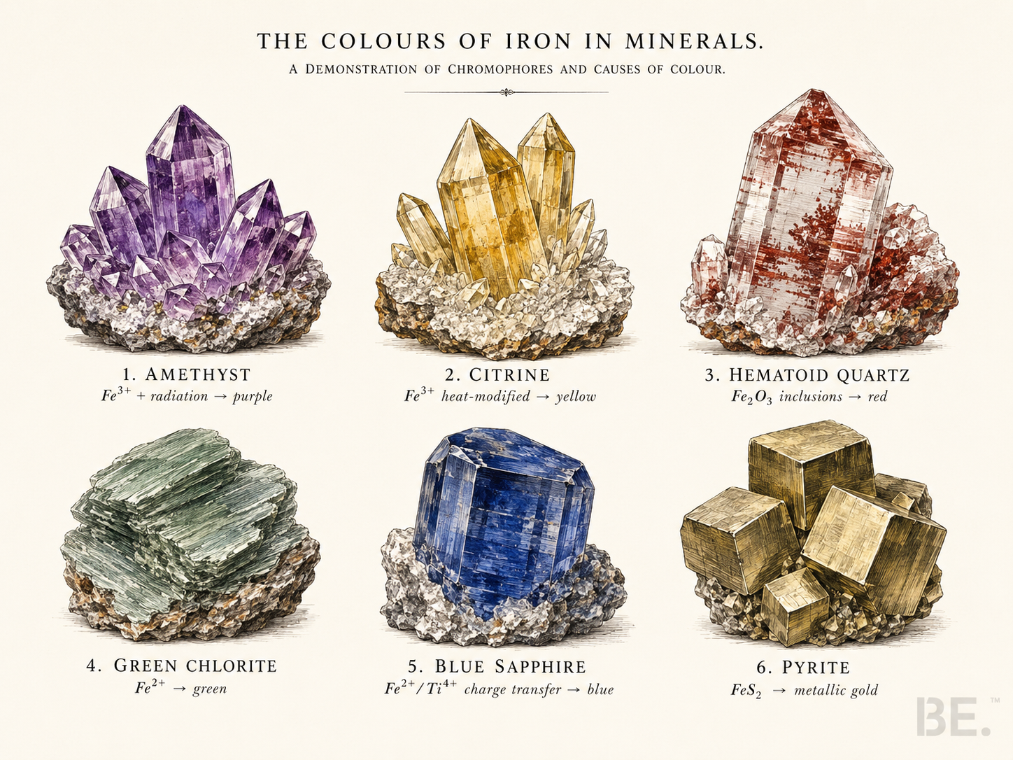
| Hematoid quartz | Fe³⁺ (as Fe₂O₃ inclusions) | Discrete hematite particles, not lattice iron | Red, orange, brown veining | Colour comes from hematite/limonite inclusions floating inside clear quartz; not a lattice substitution. |
| Aquamarine (beryl) | Fe²⁺ (and Fe³⁺ in some) | Channel sites and octahedral Al sites | Sky blue to sea-green | Fe²⁺ broad NIR absorption + occasional Fe²⁺-Fe³⁺ IVCT in the channel. Heat treatment shifts the ratio toward blue. |
| Amethyst (quartz) | Fe³⁺ (after ionising radiation) | Fe³⁺ substituting for Si⁴⁺ + a hole colour centre | Purple to violet | Natural radiation removes an electron, leaving a stable Fe-related hole centre that absorbs in yellow-green; violet survives. |
| Citrine (quartz) | Fe³⁺ (heat-modified or natural) | Fe³⁺ as Fe(OH) or Fe₂O₃-like clusters in lattice | Yellow to orange | Heating amethyst converts the hole-centre Fe³⁺ to a different cluster geometry; the absorption shifts into the violet end and yellow transmits. |
| Peridot (olivine) | Fe²⁺ | Octahedral M1/M2 sites (with Mg) | Yellow-green to olive | Fe²⁺ in olivine’s distorted octahedron absorbs at red and blue ends, transmitting in the green-yellow band. |
| Prehnite (with epidote inclusions) | Fe³⁺ | Octahedral Al/Fe site | Yellow-green to apple green | Fe³⁺ in the prehnite lattice — and epidote needle inclusions — combine to give the cool herbal green. |
| Hematite | Fe³⁺ | Octahedral, Fe₂O₃ structure | Metallic grey to red streak | Strong O→Fe charge transfer absorbs visible light broadly; the surface looks metallic, the powder is red. |
| Tiger’s eye | Fe³⁺ (limonite-stained) | Replaced crocidolite fibres + iron oxide film | Gold to brown chatoyancy | Limonite (Fe oxyhydroxide) coats fibrous quartz pseudomorphs; reflectance + iron stain give the warm sheen. |
| Pyrite | Fe²⁺ (in FeS₂) | Octahedral in iron disulfide | Brassy gold (metallic) | Not a colour-centre transition. Pyrite is a metallic semiconductor; the gold reflectance comes from band structure, not d-d. |
What the table is really saying
Read down the second column: Fe²⁺, Fe²⁺, Fe³⁺, Fe³⁺, Fe²⁺, Fe³⁺, Fe³⁺. The visible result column does not follow it. Iron is not the dial; iron is the input. The dial is the host structure plus the oxidation state plus the local geometry. That is why a single element can travel through a catalogue and look red here, blue there, yellow elsewhere — and why “iron = red” is one of the more persistent mineralogical misreadings.
It also explains why heat treatment, irradiation, and tectonic burial can shift a stone’s colour without changing its iron content. Amethyst becomes citrine on heating because the Fe-related colour centre is restructured, not because iron leaves. Aquamarine deepens after thermal treatment because Fe³⁺ is reduced toward Fe²⁺. The colour follows the chemistry of the centre, not the bulk count of iron atoms.
How BE. reads iron in a strand
BE.’s grading framework treats colour as a material fact, not a metaphor. When you look at a garnet strand and a hematoid strand side by side, both read as “iron stones.” Materially, they are not the same: garnet’s red is a structural fact about the dodecahedral site holding Fe²⁺ in an aluminosilicate cage; hematoid’s red is a particle suspension — flakes of Fe₂O₃ in an otherwise transparent quartz body. Both legitimate. Different mechanisms.
The BE. Crystal 4T Grading System reads transparency, tone, texture and treasure separately, so two iron stones do not get collapsed into one category. The BE. Geological Codex documents the formation path and inclusion type for each piece so that the colour story can be told as material, not magic.
Frequently asked questions
Q1.If iron has no fixed colour, why is rust red?
Rust is iron oxide — Fe³⁺ in oxide and hydroxide form. The bulk solid has a strong O→Fe charge transfer that absorbs across the blue and green band, leaving red-brown reflectance. That is one specific structural environment for iron. In a different host lattice (beryl, olivine, quartz, sapphire), the same Fe³⁺ behaves differently.
Q2.Is hematoid quartz the same kind of red as garnet?
No. Garnet’s red is intrinsic — the iron is locked into the crystal lattice and produces the colour through electron transitions. Hematoid quartz is clear quartz with hematite inclusions; the red is a particle suspension, not a lattice transition. Visually similar in some hands, structurally very different.
Q3.Why does amethyst look purple if iron is the colouring agent?
Amethyst’s colour requires three things together: trace Fe³⁺ substituting for Si⁴⁺, ionising radiation to remove an electron, and a stable hole colour centre that absorbs yellow-green wavelengths. The transmitted wavelengths sit in the violet band. Heat the stone enough and the centre restructures into a different geometry — that is citrine.
Q4.Can you tell iron oxidation state by eye?
Sometimes, with experience. Deep red typically signals Fe²⁺ in a 6- or 8-coordinated site; rusty orange-brown signals Fe³⁺ oxide forms; sky-blue often signals Fe²⁺ with possible IVCT; yellow-green can be either, depending on the host. Definitive readings require spectroscopy.
Q5.Does iron content matter for stone quality?
Quantity matters less than where the iron sits. A small amount of iron in the right site produces saturated colour; a large amount in the wrong site can produce a muddy result. Grading is about structural fit, not bulk concentration.
References
- Mindat — Garnet group — site geometry and almandine end-member chemistry.
- Mindat — Beryl — channel iron substitution in aquamarine.
- Burns, R.G. (1993). Mineralogical Applications of Crystal Field Theory, 2nd ed., Cambridge University Press — the standard treatment of iron d-d transitions and IVCT.
- Nassau, K. (1983). The Physics and Chemistry of Color: The Fifteen Causes of Color, Wiley — broad framework for colour-centre and charge-transfer mechanisms.
- BE. Crystal 4T Grading System — the four observable axes used to read a strand.
- BE. Geological Codex — material-level reference for the stones in the BE. catalogue.